Neuro-Anatomy
Structure/location in the brain
Brain Location
Different specific sites in your brain are responsible for or involved in specific brain functions. Therefore, training different locations of your brain will give you different outcomes (based on the functions of the underlying brain).
Vision, hearing, and other senses
The very back of your brain (occipital lobes) contain the primary visual cortex and the secondary visual cortex. These areas are entirely responsible for creating/processing vision from signals that come from your eyes. It turns out you don’t actually “see” with your eyes, but you “see” with your brain in the visual cortex. Of course you need your eyes to see because your eyes take in light through your lens and direct that light onto your retina, where the cells (rods and cones) convert the light into electrical activity that passes through the optic nerves (the 2nd cranial nerve), to your visual cortex where vision is created/processed. Then through connections with other parts of your brain, you can interpret the meaning of these vision signals and decide how you should react to this newly created vision. The same thing is true for all of your senses. You don’t actually create/process hearing with your ears, your brain does that in a specific location in the temporal lobe called the auditory cortex. Sound waves move your eardrum, and that causes tiny inner ear “ossicles” to create electrical signals that travel through the auditory nerve (the 8th cranial nerve) to your auditory brain cortex where hearing is created/processed. The same is true for smell. The chemical molecules that enter your nose stimulate electrical signals that travel through the olfactory nerve (1st cranial nerve) to your olfactory brain cortex. Of course, you can probably guess that dogs have a much larger portion of their brains devoted to smell than humans do.
Body Sensations and Pain: The Pain Network
In the same way as vision, hearing, and smell, your sense of touch or sensation from your entire body creates electrical signals at your nerve endings, which then travel through your peripheral nerves, your spinal cord, and then directly to your brain for creating/processing sensations, including pain. The most important locations for pain are the thalamus, somatosensory cortex (SSC), insular cortex and anterior cingulate cortex (ACC). These brain locations (combined with a few other specific locations) make up a complex web of connections between areas of your brain that create/process pain and suffering. These specific brain areas are collectively called the Pain Network. There are actually 3 distinct pain pathways that send signals to distinct parts of the Pain Network, where pain is created/processed. These are known as the: 1) primary pain pathway, 2) suffering pain pathway, and 3) pain inhibition pathway. As these names suggest, if you have too much activity in the pain suffering pathway and too little activity in the pain inhibition pathway, you will almost certainly suffer from chronic pain, whether it be chronic migraines, chronic face pain, chronic neck/shoulder/back pain, abdominal pain without a structural cause (IBS), chronic pelvic pain, or extremity pain (such as CRPS or RSD).
Location of Pain in your body: The Homunculus
The location of your chronic pain with excessive suffering also depends on what exact location of your cerebral Pain Network has abnormal electro physiological (EP) activity. You see, your brain has every part of your body mapped out in very specific locations within the Pain Network. If we stimulate specific parts of your SSC In the pain network, (for example the face area of your right SSC), you will feel that stimulated sensation in the exact part of your body that was activated/stimulated (in this example in your left face). This stimulation is sometimes performed during brain surgery (for example when removing a brain tumor) to help us neurosurgeons avoid the areas of your brain that control movement and sensation for each of your body parts.
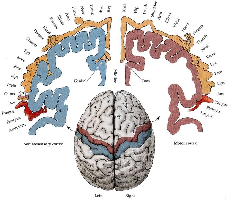
The very top of the SSC and motor cortex create/control the sensations and movements in your feet. Likewise, the bottom part of the SSC and motor cortex create/control sensations and movement of your face. Every other body part is controlled by the brain cortex between the foot and face. If you draw all the parts of your body that are controlled by each exact area of the SSC and motor cortex, you will have a funny-proportioned “little human” called a homunculus, as illustrated below. The specific part of this homunculus that has abnormal activity within the Pain Network determines where you have pain in your body. This explains why it’s possible to have severe pain in your right foot even though you don’t have a right foot because it was amputated years ago (this is known as phantom pain). Phantom pain can only be explained by the pain being created/processed in the Pain Network in your brain. Specifically, the part that controls the right foot (that portion of the brain responsible for causing pain in your right foot is still working, even though your right leg/foot is missing). It turns out, ALL pain is created/processed in your Pain Network in your brain. If you step on a nail, the nail doesn’t “cause/create” the pain (but it starts the process). The nail stimulates your nerve endings in your foot which then send electrical signals up the nerves in your leg, then up the spinal cord, then to your Pain Network within your brain, and this is where the pain is created/processed (the decision for pain is made here). Because this is true, we can perform a Brain Map that shows the abnormal activity within your brain, and then we can use this exact information to turn the intensity of the Pain Network down, so it is more tolerable or gone completely.

330 22nd Ave N.
Nashville, TN 37203
615-988-1181
Copyright 2026 Expertise Health
Location
Monday
Tuesday
Wednesday
Thursday
Friday
Hours
9:00 AM - 5:30 PM
9:00 AM - 5:30 PM
9:00 AM - 5:30 PM
9:00 AM - 5:30 PM
9:00 AM - 5:30 PM